What is ultrasound imaging?
Ultrasound is a modern, affordable, and safe method of diagnosis which helps to create an image of internal organs, soft tissues, and blood vessel walls. This method is based on the ability of sound waves to echo off various body structures.
Ultrasound is used in all areas of medicine, thanks to it being informative, painless, and having almost no contraindications.
Modern ultrasound machines can provide images of internal body structures up to 30 cm deep.
 In order to examine blood vessels, ultrasound is combined with Doppler ultrasonography, which helps to measure the velocity and direction of blood flow. The same scan, but with a color image, is called a triplex ultrasound.
Heart ultrasound, or echocardiography, helps to assess the condition of heart valves, heart muscle parameters, the size of heart chambers, the thickness of its walls, the presence of blood clots, etc. Thanks to modern equipment, it is also possible to determine the direction and speed of blood flow in heart chambers.
Ultrasound helps to:
- find structural changes in internal organs, as well as their disorders.
- monitor the changes as often as necessary, thanks to the safety of this method.
- determine the patency of important blood vessels, the condition of blood vessel walls, and blood flow disorders (narrowing, blood clots, atherosclerotic plaques, etc.).
- examine the heart condition.
- to identify the presence of pathological fluid in internal organs (abdominal cavity, pelvic cavity, organs of the thorax, joints, etc.).
- study the anatomy of the pathological area in order to carry out diagnostic or surgical intervention (paracentesis, biopsy, etc.). It is also used for navigation during surgery (e.g., during blood vessel surgeries).
- to assess the parameters and stage of pregnancy, the condition of a fetus.
Ultrasound imaging at Kivach clinic
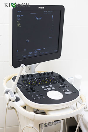
The clinic's diagnostic rooms are equipped with modern and advanced ultrasound machines that can record video.
Ultrasound advantages:
- Informativity. It helps to get more information about body structures over a shorter period of time.
- Precise diagnostic images.
- Convenience. Saved images and videos can be compared with the results of previous and future examinations.
Ultrasound is used in all areas of medicine as the most accessible and easy to use method of diagnosis.
Indications:
- To monitor a detected pathology (as a rule no less than once a year).
- Diseases of the abdominal organs.
- Kidney and bladder diseases.
- Thyroid gland diseases.
- Heart diseases, arterial hypertension.
- Blood vessel diseases, varicose and atherosclerotic diseases.
- Gynecological disorders.
- Mammary gland disorders. Apart from that, ultrasound of mammary glands is done once in every two years for women under 40 as a preventive measure (women over 40 are recommended to have a mammogram).
- Prostate diseases. Annual prostate screening is recommended for all men after 50.
- To examine the fetus during pregnancy.
What to expect during the test?
The ultrasound is performed in a special room with dim light. The patient is positioned according to the type of examination – he/she might lay down, stand, or sit.

A sonographer uses a transducer that is suitable for the examined area. A special gel is applied onto the transducer. The sonographer moves the transducer over the examined area, with the transducer pressed against a patient’s body. The image of internal organs appears on the screen. The doctor captures the size, tissue density, and details of anatomical structures.
During the process, a patient might be asked to hold his/her breath or change the position of his/her body.
The exam usually lasts for 15-30 minutes.
In the report, the doctor will describe the examined area, its size, and other calculated parameters. A doctor's consultation is required should there be an abnormality.
Some types of ultrasound exams have their distinctions.
Pelvic ultrasound is performed transvaginally using a special transducer, which is inserted into a woman's vagina. The patient lays down with the legs slightly spread out.
Prostate ultrasound is performed transrectally, with the transducer being inserted into the rectum about 5 cm deep. The patient lays down on his side with his knees bent.
Contraindications
There are no absolute contraindications for this method.
The exam is not performed if it is impossible to render an image of internal organs as a result of:
- Skin damage or pain in the examined area, which makes it impossible for the transducer to contact the skin.
- Excess subcutaneous fat and significant bloating make it harder to render an image.
- Urinary incontinence for exams which require a full bladder.
- Acute vaginal and cervical infectious diseases is a contraindication for a transvaginal exam.
- Acute rectal disorders, exacerbation of hemorrhoids, anal fissures are contraindications for transrectal examination.
Question-answer
- Is the exam safe?
-
Yes, the exam is safe.
- Is the exam painful?
-
The exam is painless.
In some cases, the pressure from the transducer might cause discomfort. There might be a minor discomfort at the beginning of the exam when cold gel contacts the skin. - How to prepare for the exam?
-
Only some types of ultrasound exams require preparation.
- Are complications possible?
-
There are no complications.
- What ensures that the exam is successful?
-
- Qualified medical specialists with extensive medical experience.
- Advanced equipment.
- Compliance with the standards of medical care.

